

 QuranGPT
The 576-Megapixel Marvel: The Neuroscience, Optics, and Divine Architecture of the Human Eye
An exhaustive scientific and Quranic exploration into the 576-megapixel optical architecture of the human eye, photoreceptor biochemistry, and divine creation.
The Holy Quran Neuroscience & Spiritual Health Editorial
Author
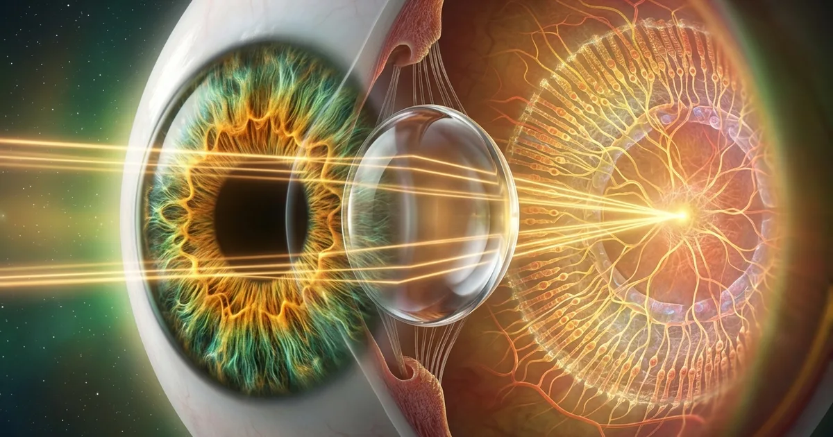
Among the trillions of biological structures fashioned in the living universe, none presents a more astonishing synthesis of optical physics, quantum biochemistry, and neural computation than the human eye. Operating as an uninterrupted, self-cleaning, autofocusing biological camera that consumes less than two watts of metabolic energy, the human visual apparatus perceives reality with a fidelity that surpasses the most advanced semiconductor image sensors manufactured by human engineering.
For centuries, natural philosophers, optical engineers, and neurobiologists have attempted to quantify the limits of human visual perception. When translated into modern digital sensor metrics, the composite resolving power of the human visual system equates to an astonishing 576 megapixels. Yet this staggering resolution represents only a single dimension of its brilliance. The human eye functions not as a static digital sensor capturing isolated frames, but as a dynamic quantum-mechanical photon detector coupled to a massively parallel neural supercomputer—the brain's visual cortex.
In this exhaustive investigation, we explore the optical physics, microscopic photoreceptor biochemistry, mathematical resolution models, and the profound theological dimensions underlying the creation of human vision (Al-Basar), demonstrating how modern biophysics affirms the timeless eloquence of the Holy Quran.
1. Executive Summary: The Biological Optical Matrix
To understand the magnitude of human vision, one must compare its physiological parameters against state-of-the-art commercial camera sensors:
| Optical & Anatomical Parameter | Biological Human Eye (Homo sapiens) | Flagship Medium-Format Digital Camera (2026) |
|---|---|---|
| Composite Visual Resolution | 576 Megapixels (Integrated Binocular FOV) | 100 to 150 Megapixels |
| Instantaneous Foveal Resolution | 7 to 10 Megapixels (Central 2° Arc) | 61 to 100 Megapixels (Uniform across sensor) |
| Total Photoreceptor Count | ~126 Million (120M Rods + 6M Cones) | 61 to 150 Million Photodiodes |
| Dynamic Contrast Range | 1,000,000 : 1 (20 to 24 F-Stops HDR) | 14 to 16 F-Stops |
| Focal Length & Aperture Range | f/2.1 (Night) to f/8.3 (Bright Sunlight) | Lens dependent (e.g., f/1.4 to f/22) |
| Optical Refractive Power | +58 to +65 Diopters (Cornea + Lens) | Varies with multi-element glass groups |
| Latency / Refresh Rate | Continuous analog integration (~100–120 Hz equivalent) | 30 to 120 FPS Electronic Shutter |
| Data Transmission Trunk | 1.2 Million Myelinated Axons (Optic Nerve) | PCIe 4.0 / USB 4.0 Fiber (~40 Gbps) |
| Power Consumption | < 2 Watts metabolic energy | 15 to 35 Watts (Battery + Active Cooler) |
2. The Mathematics Behind the 576-Megapixel Calculation
The widely cited 576-megapixel benchmark was formulated by astrophysicist, planetary scientist, and optical researcher Dr. Roger N. Clark (Visual Acuity of the Human Eye and Digital Camera Equivalents). To appreciate how this number is derived, we must understand the fundamental difference between a silicon sensor and biological human vision.
A. Visual Acuity and Arcminutes
Human visual acuity is measured by the ability to distinguish two separate points of light separated by an angular distance. In an individual with standard 20/20 (or 6/6) Snellen vision, the maximum resolving power of the eye is 1 arcminute (1/60th of a degree). However, under optimal high-contrast lighting conditions, the densely packed cone photoreceptors in the human fovea can resolve details down to 0.3 to 0.5 arcminutes (0.005 to 0.008 degrees).
Visual Angle Calculation:
1 Degree (°) = 60 Arcminutes (')
0.5 Arcminute = 1/120th of a Degree (Standard Optical Acuity Limit)
0.3 Arcminute = 1/200th of a Degree (Peak Foveal Acuity Limit)
B. The Spatial Field of View (FOV)
When keeping both eyes open and gazing forward, the binocular human field of view spans approximately 120 degrees horizontally by 120 degrees vertically (with peripheral awareness extending to nearly 180 degrees horizontally).
To calculate the number of independent optical sampling points (pixels) required to populate a 120° by 120° field of view at a maximum resolving capability of 0.3 arcminutes per pixel:
Horizontal Pixel Count = (120 degrees × 60 arcminutes/degree) / 0.3 arcminutes/pixel
                       = 7,200 / 0.3
                       = 24,000 pixels

Vertical Pixel Count   = (120 degrees × 60 arcminutes/degree) / 0.3 arcminutes/pixel
                       = 7,200 / 0.3
                       = 24,000 pixels

Total Equivalent Pixels = 24,000 × 24,000
                        = 576,000,000 pixels (576 Megapixels)
C. Foveal Gaze vs. Saccadic Integration
A digital camera captures its entire frame uniformly at full resolution across the whole sensor. If the human eye attempted to capture 576 megapixels uniformly in a single instant, the optic nerve would need to be as thick as an arm, and the brain would suffer severe metabolic overload.
Instead, the Creator engineered a vastly superior efficiency architecture:
- The Instantaneous Core (Fovea Centralis): High-density color vision is concentrated in a tiny 1.5-millimeter zone of the retina called the fovea, spanning only 2 degrees of arc (equivalent to the width of your thumbnail held at arm's length). In this tiny core, the resolution is approximately 7 to 10 megapixels.
- The Peripheral Field: Surrounding the fovea, photoreceptor density drops dramatically, prioritizing motion detection, luminance contrast, and spatial orientation over sharp detail.
- Rapid Saccadic Scanning: The brain directs the eyes in rapid, subconscious micro-movements called saccades (3 to 4 saccades per second), darting across the visual scene.
- Cortical Buffer Synthesis: The visual cortex seamlessly stitches these high-resolution foveal samples into a continuous, crystal-clear 576-megapixel mental image buffer in real time.
3. Anatomical Architecture: The Tri-Partite Refractive System
The physical eye is a masterclass in organic engineering, consisting of three primary refractive media working in perfect optical harmony:
Incoming Photons ──► Tear Film & Cornea (+43D) ──► Aqueous Humor & Pupil ──► Crystalline Lens (+15D to +20D) ──► Vitreous Body ──► Inverted Retinal Plane
1. The Cornea: Aspheric Precision Refraction
The front transparent window of the eye, the cornea, provides approximately 70% to 75% (+40 to +44 Diopters) of the eye's total optical focusing power. Unlike human-made glass lenses which suffer from spherical aberration (where light rays passing through the lens edge focus at a different point than central rays), the cornea is naturally aspheric—it flattens progressively toward the periphery, perfectly neutralizing spherical aberration across all visible wavelengths.
2. The Dynamic Iris and Pupil: Biological Aperture Control
Surrounding the pupil is the iris, a colored diaphragm composed of two opposing muscle groups: the sphincter pupillae (circular constrictor muscles operated by parasympathetic nerves) and the dilator pupillae (radial dilator muscles operated by sympathetic nerves). In less than 200 milliseconds, the pupil modulates its aperture from 2.0 mm (in bright desert sun) to 8.0 mm (in deep twilight), varying light intake by a factor of 16 without introducing chromatic distortion.
3. The Crystalline Lens: Variable-Curvature Autofocus
Positioned directly behind the iris is the biconvex crystalline lens. Composed of specialized transparent proteins called crystallins, the lens achieves what no mechanical camera lens has ever duplicated: continuous variable-curvature deformation. Connected to the circular ciliary muscle via thousands of micro-filaments (zonules of Zinn), the lens dynamically alters its thickness in milliseconds:
- Distant Vision: The ciliary muscle relaxes, zonules pull tight, flattening the lens for optical infinity.
- Near Vision (Accommodation): The ciliary muscle contracts, releasing zonular tension, allowing the elastic lens capsule to round up, adding +15 to +20 diopters of refractive power to read fine text held inches from the face.
4. The Retinal Mosaic: 126 Million Photoreceptor Cells
At the posterior pole of the ocular sphere lies the retina, a paper-thin layer of specialized neural tissue only 0.2 to 0.5 millimeters thick. Despite its microscopic delicacy, the retina houses approximately 126 million light-sensitive photoreceptor cells organized into two distinct classes:

A. Rod Photoreceptors (~120 Million Cells)
- Primary Function: Scotopic vision (night vision, low-light detection, monochrome contrast).
- Quantum Sensitivity: Capable of generating an electrical signal from a single photon of light.
- Distribution: Dense across the peripheral retina; completely absent in the center of the fovea.
- Photopigment: Rhodopsin (peak spectral absorption at 498 nm, blue-green light).
B. Cone Photoreceptors (~6 to 7 Million Cells)
- Primary Function: Photopic vision (daylight vision, high-acuity fine detail, trichromatic color perception).
- Distribution: Extremely concentrated within the fovea centralis (densities reaching 200,000 cones per square millimeter).
- The Three Chromatic Channels:
- S-Cones (Short-wavelength / Blue): Peak absorption at ~420 nm (constituting ~5-10% of cones).
- M-Cones (Medium-wavelength / Green): Peak absorption at ~534 nm.
- L-Cones (Long-wavelength / Red): Peak absorption at ~564 nm.
By comparing the differential excitation ratios across these three cone channels, the brain discriminates over 10 million distinct color gradations across the electromagnetic spectrum (380 nm to 740 nm).
5. Phototransduction: The Sub-Millisecond Quantum Chemistry of Vision
How does an incoming electromagnetic wave (a photon of light) transform into a conscious thought inside the brain? The biochemical mechanism, known as the phototransduction cascade, is one of the most elegant molecular cascades in all of cellular biochemistry:
Photon Absorption (hν) ──► 11-cis-retinal isomerizes to All-trans-retinal ──► Metarhodopsin II ──► G-Protein Transducin Activation ──► PDE6 Hydrolyzes cGMP ──► Sodium/Calcium Channels Close ──► Photoreceptor Hyperpolarization (-70 mV) ──► Decreased Glutamate Release ──► Bipolar & Ganglion Action Potentials
- The Molecular Antenna: Embedded within the membranous discs of rod cells are millions of Rhodopsin molecules. At the center of each rhodopsin protein sits a molecule of 11-cis-retinal (derived from Vitamin A).
- Light-Induced Isomerization: When a single photon strikes the 11-cis-retinal molecule, its kinked hydrocarbon chain snaps into a straight all-trans-retinal shape within 200 femtoseconds (0.0000000000002 seconds)—one of the fastest known chemical reactions in nature.
- Biochemical Amplification: This shape change converts rhodopsin into Metarhodopsin II, which activates hundreds of molecules of the G-protein Transducin. Each transducin molecule activates Phosphodiesterase-6 (PDE6), which rapidly hydrolyzes thousands of cyclic GMP (cGMP) molecules.
- Electrical Hyperpolarization: The depletion of cGMP causes cGMP-gated cation channels in the cell membrane to close, stopping the influx of sodium (Na⁺) and calcium (Ca²⁺) ions. The cell's electrical potential drops from -40 mV to -70 mV (hyperpolarization).
- Signal Transmission: This change in voltage modulates neurotransmitter release (glutamate) across the synaptic cleft to bipolar cells, which in turn fire action potentials down the 1.2 million fibers of the optic nerve directly to the visual processing centers of the brain.
6. Divine Teleology: The Quranic Dimension of Sight (Al-Basar)
In the Holy Quran, the faculties of perception are repeatedly cited as among the greatest signs of Divine creation, wisdom, and benevolence. The Arabic term for sight—الْبَصَر (Al-Basar) and its plural الْأَبْصَار (Al-Absar)—occurs dozens of times throughout the Quranic text, constantly juxtaposed with hearing (As-Sam') and cognitive reflection (Al-Af'idah).
A. The Primacy of Sensory Bestowal
In Surah Al-Mulk, the Creator reminds humanity of the miraculous origin of these sensory organs:
قُلْ هُوَ الَّذِي أَنشَأَكُمْ وَجَعَلَ لَكُمُ السَّمْعَ وَالْأَبْصَارَ وَالْأَفْئِدَةَ ۖ قَلِيلًا مَّا تَشْكُرُونَ
"Say, 'It is He who produced you and made for you hearing and vision and hearts (intellect); little are you grateful.'"
— Surah Al-Mulk (67:23)
Tafsir Insight: The great classical exegete Ibn Kathir (1301–1373 CE) notes that in this verse, the Almighty highlights the specific bestowal of Al-Absar (the visual faculties) as an unmatched blessing of discernment, enabling human beings to perceive light, color, spatial harmony, and the cosmic signs spread across the horizon.
B. The Dual Structural Gift
In Surah Al-Balad, the specific physical endowment of two stereoscopic eyes is highlighted:
أَلَمْ نَجْعَل لَّهُ عَيْنَيْنِ • وَلِسَانًا وَشَفَتَيْنِ
"Have We not made for him two eyes? And a tongue and two lips?"
— Surah Al-Balad (90:8–9)
Scientific Reflection: The Quran explicitly emphasizes "two eyes" ('aynayn). Stereoscopic binocular vision requires two horizontally separated optical sensors whose optical axes converge with sub-millimeter precision. Without two coordinated eyes, depth perception, three-dimensional spatial parallax, and stereoscopic distance calculation would be physically impossible.
C. The Inverted Order of Sensory Development
Modern embryology has demonstrated that during fetal gestation in the womb, the auditory system develops and becomes functional before the visual system. The inner ear cochlea and auditory nerve fully form by the 22nd to 24th week of gestation, whereas the eyes remain fused shut until approximately the 26th week, and functional retinal neural processing is only calibrated post-birth upon exposure to photon stimulation.
Remarkably, throughout the Quran, whenever hearing and sight are mentioned together, hearing (As-Sam') always precedes sight (Al-Absar):
وَاللَّهُ أَخْرَجَكُم مِّن بُطُونِ أُمَّهَاتِكُمْ لَا تَعْلَمُونَ شَيْئًا وَجَعَلَ لَكُمُ السَّمْعَ وَالْأَبْصَارَ وَالْأَفْئِدَةَ ۙ لَعَلَّكُمْ تَشْكُرُونَ
"And Allah brought you out from the wombs of your mothers while you knew nothing, and He gave you hearing and sight and intellect that perhaps you might be grateful."
— Surah An-Nahl (16:78)
7. Dynamic Range & Low-Light Sensitivity: Biological HDR
Human-made digital cameras struggle immensely in scenes containing both brilliant highlights and deep shadows (such as standing inside a dark room while looking out a sunlit window). A standard digital sensor achieves roughly 10 to 14 stops of dynamic range.
The human visual system, by contrast, commands an astonishing dynamic range of 20 to 24 stops (a contrast ratio exceeding 1,000,000 : 1):
Bright Sunlight on Snow (~100,000 Lux) ◄────────────────────────► Starlit Moonless Night (~0.0001 Lux)
              [A dynamic operating span of 1 Billion to 1 in light intensity]
How the Eye Achieves Biological HDR:
- Dual Sensor Architecture (Duplicity Theory): The retina operates two distinct biological sensor systems in parallel: Cones handle bright lighting (Photopic), while Rods take over in darkness (Scotopic). In intermediate lighting (Mesopic, such as twilight), both systems blend seamlessly.
- Pupillary Light Reflex: The iris dilates and constricts to instantly adjust light flux by a factor of 16.
- Photopigment Bleaching & Regeneration: In intense illumination, excess rhodopsin bleaches into retinal and opsin, desensitizing the rods to prevent signal saturation. In darkness, the retinal pigment epithelium (RPE) continuously regenerates fresh 11-cis-retinal, multiplying low-light sensitivity by up to 100,000-fold over a 30-minute dark-adaptation period.
- Neural Gain Control: Retinal ganglion cells dynamically adjust their firing thresholds, shifting baseline exposure values locally across different regions of the visual field.
8. The Brain as the Neural GPU: Visual Cortex Processing
The eye does not "see"—the eye gathers and pre-filters optical data; the brain sees. The optic nerve transmits raw visual telemetry at an estimated bandwidth of 10 megabits per second into the Lateral Geniculate Nucleus (LGN) of the thalamus, which routes the data into the Primary Visual Cortex (V1 / Brodmann Area 17) at the back of the occipital lobe.
Retina (126M Sensors) ──► Optic Nerve (1.2M Lines) ──► Thalamus (LGN Router) ──► Primary Visual Cortex V1 (Edge Detection) ──► V2/V3 (Form & Depth) ──► V4 (Color Processing) ──► V5/MT (Motion Tracking) ──► Conscious Awareness
Key Neural Processing Feats:
- Inverted Image Rectification: The cornea and lens focus an upside-down, mirror-reversed image onto the curved retina. The visual cortex re-maps and rectifies this inverted spatial projection into an upright, seamless 3D mental model of reality.
- Blind Spot Inpainting: Where the optic nerve exits the eyeball (the optic disc), there are zero photoreceptors, creating an absolute blind spot in each eye. The visual cortex uses contextual AI-like interpolation to "inpaint" and fill the blind spot with matching texture and color so that humans never perceive a black hole in their field of vision.
- Saccadic Suppression: When the eyes dart from one point to another at speeds exceeding 900 degrees per second, the brain temporarily suspends visual processing for 50 milliseconds (saccadic masking). This prevents the dizzying motion blur that would otherwise occur every time we look around.
9. Classical Islamic Optics: Ibn al-Haytham’s Scientific Revolution
The foundational physics governing our understanding of the eye and lenses was pioneered by the great 11th-century Islamic polymath Al-Hasan Ibn al-Haytham (Alhazen, 965–1040 CE) in Cairo, Egypt.
       CLASSICAL GREEK VIEW (Emission)                  IBN AL-HAYTHAM'S BREAKTHROUGH (Intromission)
       [Euclid & Ptolemy]                               [Kitab al-Manazir - 1021 CE]

       Eye ════════(Emits Visual Rays)══════► Object     Light Source ────► Object ────(Reflected Rays)────► Eye
       [INCORRECT: Eye radiates light]                   [CORRECT: Eye receives reflected photons]
In his monumental seven-volume masterpiece, Kitāb al-Manāẓir (The Book of Optics), Ibn al-Haytham:
- Dismantled Greek Emission Theory: Proved experimentally that visual rays do not originate from the eye (refuting Euclid and Ptolemy), but rather light reflects off objects and enters the eye (Intromission Theory).
- Discovered the Principle of the Camera Obscura (Al-Bayt Al-Muthlim): Demonstrated how light rays passing through a tiny pinhole form an inverted image on a darkened surface—the exact physical principle upon which both the human eye and modern digital cameras are based.
- Pioneered Anatomical Optics: Provided the first accurate scientific diagrams illustrating the cornea, aqueous humor, crystalline lens, vitreous humor, and the optic nerve pathway to the brain.
10. Technical Comparison: Digital Camera vs. The Human Eye
| Feature | Human Eye | Premium Mirrorless Digital Camera |
|---|---|---|
| Total Resolution | 576 Megapixels (Composite FOV) | 24 to 61 Megapixels |
| Sensor Shape | Spherical / Curved Retina (Zero edge distortion) | Flat Rectangular Silicon Wafer |
| Edge Aberrations | Zero spherical / coma distortion (Curved surface matches focal plane) | Requires 10 to 18 glass elements to correct flat-field curvature |
| Autofocus Mechanism | Biological muscle deformation of flexible lens | Mechanical motors moving heavy glass elements |
| Autofocus Speed | ~350 milliseconds | 100 to 400 milliseconds |
| Operating Power | ~1 to 2 Watts (ATP glucose consumption) | 10 to 30 Watts (Lithium-ion battery) |
| Self-Maintenance | Continuous biological cellular renewal & tears | Requires manual lens cleaning & sensor swab |
| Lifespan | 80 to 100+ years of continuous 24/7 operation | ~200,000 to 500,000 mechanical shutter actuations |
11. Frequently Asked Questions (FAQ)
Q1: Is the human eye really 576 megapixels?
Yes, when measured as the total resolving capacity of the brain-eye system across the complete binocular field of view (120° × 120°) at standard foveal acuity (0.3 to 0.5 arcminutes per pixel). While a single static gaze registers about 7 to 10 megapixels in the central fovea, rapid subconscious micro-saccades (3 to 4 per second) allow the visual cortex to composite a seamless 576-megapixel mental image.
Q2: Why don't we notice our blind spot?
The blind spot occurs at the optic disc, where the 1.2 million nerve fibers leave the eye to form the optic nerve. We do not notice it because our two eyes have overlapping visual fields that cover each other's blind spots, and the visual cortex dynamically "inpaints" the missing region using surrounding background patterns, colors, and textures.
Q3: How does the human eye achieve high dynamic range (HDR)?
The eye achieves up to 24 stops of dynamic range through a multi-tiered biological adaptation system: pupillary constriction/dilation, dual photoreceptor channels (rods for darkness, cones for daylight), biochemical regeneration of rhodopsin, and neural gain adjustments in retinal ganglion cells.
Q4: What is the significance of "Hearing before Seeing" in the Quran?
In virtually every Quranic passage where sensory faculties are listed (e.g., Surah An-Nahl 16:78, Surah Al-Mulk 67:23), hearing (As-Sam') is mentioned prior to sight (Al-Absar). Modern developmental embryology confirms that the human auditory system develops, matures, and becomes functional in the womb weeks before the visual system is formed.
Q5: How many colors can the human eye differentiate?
Through the combinatorial processing of three cone types (Red/L-cones, Green/M-cones, Blue/S-cones) and subsequent opponent-process neural pathways in the brain, the human visual system can distinguish approximately 10 million distinct colors.
Q6: Who was the first Muslim scientist to explain how the eye works?
Al-Hasan Ibn al-Haytham (Alhazen, 965–1040 CE) revolutionized optics in his seminal work Kitāb al-Manāẓir. He proved that vision occurs when reflected light enters the eye (intromission theory), disproving Greek theories that the eye projected light, and laid the mathematical foundation for modern photography and ophthalmology.
12. Conclusion: A Masterpiece of Divine Craftsmanship
When we examine the human eye through the converging lenses of optical physics, neurobiology, and quantum photochemistry, the notion that such an intricate, self-adjusting, 576-megapixel system arose through unguided, blind happenstance defies every principle of statistical probability and thermodynamic logic.
From the aspheric optical clarity of the cornea and the sub-millisecond quantum isomerization of 11-cis-retinal, to the 1.2-million-fiber optic cable and the cognitive synthesis of the visual cortex, the eye stands as an irrefutable testament to the precision, wisdom, and majesty of Al-Bāri' (The Originator) and Al-Muṣawwir (The Supreme Fashioner).
هَٰذَا خَلْقُ اللَّهِ فَأَرُونِي مَاذَا خَلَقَ الَّذِينَ مِن دُونِهِ ۚ بَلِ الظَّالِمُونَ فِي ضَلَالٍ مُّبِينٍ
"This is the creation of Allah. So show Me what those other than Him have created. Rather, the wrongdoers are in clear error."
— Surah Luqman (31:11)
Summary Checklist
- ✅ 576-Megapixel Resolving Power: Formulated by Dr. Roger N. Clark, representing the human brain's seamless saccadic integration over a 120° field of view.
- ✅ 126 Million Photoreceptor Matrix: 120 million rods for extreme low-light quantum sensitivity and 6 million cones for high-acuity trichromatic color vision.
- ✅ Dynamic 24-Stop HDR Range: Seamlessly spans from 0.0001 lux (starlit night) to 100,000 lux (desert sunlight).
- ✅ Tri-Partite Optical Alignment: Cornea (+43D), dynamic iris aperture (f/2.1 to f/8.3), and deformable crystalline lens (+15D to +20D).
- ✅ Sub-Millisecond Quantum Transduction: 11-cis-retinal snaps into all-trans-retinal in 200 femtoseconds upon photon impact.
- ✅ Quranic Eloquence: Affirms Al-Basar as a profound divine blessing and mirrors embryological sensory developmental chronology.